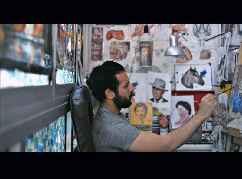
In medical schools, students begin their studies with anatomy, often going to the morgue for practical lessons on cadavers designated for this purpose. Due to the overcrowding of medical students, one veterinary medicine student in Egypt decided to help his peers by providing illustrative drawings depicting various anatomical positions of both human and animal bodies.
Egyptian student Ashraf Rafai, from Tanta in the Gharbia Governorate in northern Egypt, told "Al Arabiya.net" that he has loved drawing and art since childhood. He added that during his high school studies, he was fascinated with biology and was impressed by the simple illustrative drawings in textbooks that served as helpful study aids.
After enrolling in veterinary medicine, Rafai began studying subjects such as physiology, histology, and anatomy. He found anatomy to be the most challenging subject because it relies on direct study in the morgue and viewing cadavers. Consequently, he decided to use drawing to portray anatomical images of animals, employing renowned references like Netter, Snell, and Gray to accurately match the true colors of anatomical organs in both animals and humans.
Rafai presented his drawings to students in medical colleges through specialized libraries, and they received a great deal of interest. Students from medical, dental, and veterinary colleges began to utilize his work. He expressed happiness about this and is currently striving to learn sculpture and create specialized molds for medical anatomical models, as well as develop his drawings to be available digitally and on CDs, akin to current global references, featuring detailed explanations.
The Egyptian student dreams of creating a global reference and veterinary medical atlas for animals under his name, as there are very few medical illustrations, especially in veterinary medicine, compared to human medicine, attributed to the variety of animal species.


