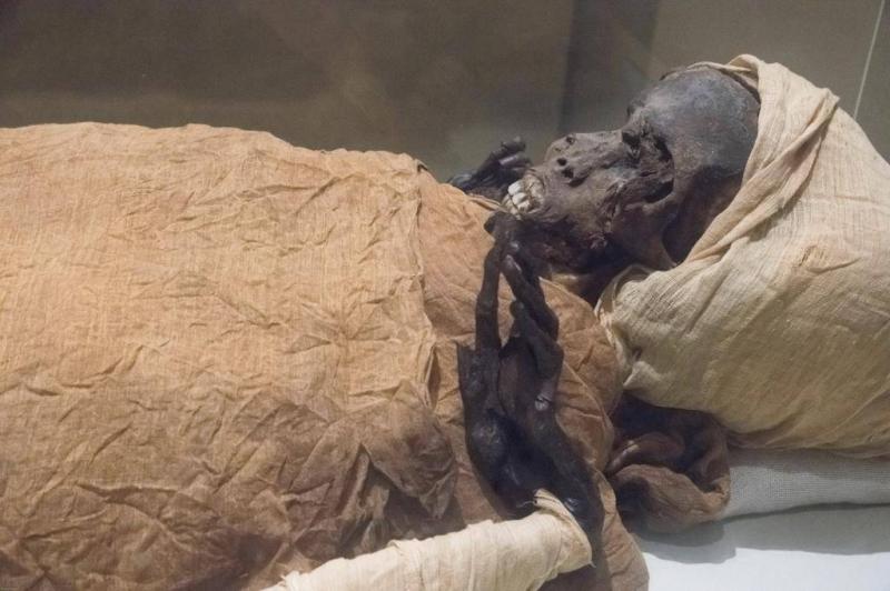
Recently, a group of researchers conducted examinations using computed tomography (CT) on the mummified remains of an ancient Egyptian pharaoh named Seqenenre Tao II, who ruled southern Egypt during the Hyksos invasion from 1650 to 1550 BC and was killed while attempting to liberate his country from foreign occupation.
The mummified remains were discovered and studied for the first time in the 1880s, and since then, scientists have questioned the precise nature of pharaoh Seqenenre Tao II’s death. In the 1960s, X-ray examinations of the mummy revealed severe head injuries without any other wounds on the mummy's body. According to archaeologists, there are two general theories about how pharaoh Seqenenre Tao II died: the first suggests he was captured in battle and subsequently executed, while the second posits that he was killed in his sleep. Previous studies noted that the condition of the mummy was poor, indicating that it may have been rapidly embalmed outside of the royal embalming workshop.
Recently, scientists used CT imaging to study the mummified remains, leading them to discover new details regarding the injuries present on the pharaoh’s mummy. Additionally, the CT imaging revealed another interesting finding: a form of lesions previously undiscovered, which had been concealed by the embalmers within the pharaoh. Researchers stated that some injuries indicate that the pharaoh was likely captured on the battlefield, and that his hands were bound behind his back to prevent him from defending himself. Notably, researchers also discovered that the pharaoh was approximately 40 years old at the time of his death, based on the morphology revealed in the new CT images. Furthermore, the CT scans showed that embalmers used an advanced technique to conceal the head wounds beneath layers of embalming material that functioned like filler used in cosmetic surgery, leading researchers to believe that the embalming was conducted in a proper embalming facility rather than hastily as suggested by previous studies.


