



Ultrasound imaging, commonly known as ultrasound, is a frequently used method for examining the kidneys and urinary tract because it is a painless and radiation-free procedure. It assists doctors in detecting larger stones and some signs of urinary tract obstructions. Consequently, it may be one of the first tests requested by a doctor in cases of flank pain or urinary symptoms.
A frequently asked question is: Can ultrasound detect kidney sand? The answer isn't strictly yes or no. The ability of ultrasound to detect sand depends on the size and location of the deposits, as well as the quality of the examination and the doctor's expertise. Very small stones might not be easily visible, whereas larger ones are more detectable.
Can Ultrasound Detect Kidney Sand?
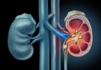
Ultrasound can reveal some deposits and stones in the kidneys, particularly when their size permits clear visibility in the image. The stone may appear as a highly echogenic area within the kidney and might sometimes be accompanied by a shadow behind it, aiding the doctor in distinguishing it.
The term 'kidney sand' often refers to very small crystals or particles that may accumulate over time to form stones. These small particles may be harder to detect by ultrasound and might not show clearly in the examination.
Therefore, the absence of sand on ultrasound does not necessarily indicate the non-existence of crystals or small stones.
What Can Ultrasound Detect in the Kidneys?
Ultrasound's role is not confined to stone detection. It can also help assess several kidney and urinary tract conditions, such as:
- Identifying some kidney stones.
- Detecting kidney pelvis dilation.
- Revealing signs of urinary tract obstruction.
- Assessing kidney size and shape.
- Detecting some cysts or structural changes.
- Evaluating the bladder and the amount of urine remaining post-micturition in certain cases.
Thus, ultrasound is considered a valuable test when there are symptoms that may indicate a urinary system issue.
Can Ultrasound Appear Normal Despite a Stone?
Yes, this can occur, especially if the stone is very small or positioned in a location challenging for ultrasound waves to clearly image.
Ultrasound may also reveal indirect signs of a stone, such as urinary tract dilation resulting from an obstruction, without the stone being visibly clear.
Therefore, doctors rely on imaging results, along with symptoms, physical examination, and urinalysis. Further tests might be needed if symptoms persist or are intense.
What's the Difference Between Kidney Sand and Kidney Stones?
People often use the term 'kidney sand' to describe small crystals or deposits formed in urine that can later accumulate into stones.
A kidney stone is a solid mass formed from substances in the urine and can vary in size, location, and composition.
Small particles may pass through the urinary tract without evident symptoms, while larger stones can cause severe pain if they move or block urine flow.
Symptoms Indicating Stones or Deposits in the Kidneys
Not all kidney stones cause symptoms, and some might be discovered incidentally during imaging for other reasons.
However, when a stone moves or causes an obstruction, symptoms may include:
- Pain on one side of the back or flank.
- Pain spreading to the lower abdomen or groin.
- Burning or pain during urination.
- Increased urge to urinate.
- Blood presence in urine.
- Nausea or vomiting.
- Cloudy or unusually smelly urine.
The severity of symptoms varies based on the stone's size, location, and the presence of obstructions or urinary tract infections.
When is a CT Scan Needed?
If symptoms are severe and ultrasound inconclusive, a physician may opt for a computed tomography (CT) scan of the kidneys and urinary tract. CT scans offer more detailed images and can detect many small stones not visible on ultrasound.
This does not mean a CT scan is necessary for everyone with flank pain; a doctor will decide the type of imaging based on symptoms, age, health status, and the likelihood of a stone or obstruction.
Can a Urine Test Detect Kidney Sand?
Urine tests can provide valuable information, but they are no substitute for imaging.
The test may reveal blood in urine, certain crystals, or signs of infection, which can help a doctor determine the potential cause of symptoms.
In some cases, further tests may be requested to identify the type of crystals or investigate underlying causes increasing the risk of stone formation.
What to Do If Ultrasound is Normal But Pain Persists?
If flank or back pain continues despite a normal ultrasound result, it is best not to presume the cause is only sand or stones.
Pain might be related to other causes, such as muscle or spinal issues, digestive problems, or urinary tract ailments.
Based on the pain's characteristics, accompanying symptoms, urinalysis, and ultrasound results, a doctor can decide if additional investigations are necessary.
When to Seek Medical Assistance Quickly?
Rapid medical evaluation is necessary for severe or sudden kidney or flank pain exhibiting particular symptoms, such as:
- Intense, unrelenting pain.
- Fever or chills.
- Persistent vomiting preventing fluid intake.
- Severe difficulty or inability to urinate.
- Noticeable blood presence in urine.
- Intense pain with urinary tract infection symptoms.
The combination of pain with fever or urinary obstruction might indicate a problem requiring urgent treatment.
How to Reduce Kidney Stone Risk?
Prevention strategies depend on the cause and type of stones, but maintaining adequate fluid intake is usually a key aspect of prevention for those without medical fluid-restricted conditions.
Doctors may advise adjusting salt and certain foods based on stone type and test results, especially for those with recurrent stones.
It's not advisable to follow strict diets or eliminate entire food groups without knowing the stone type and the underlying reasons for their formation.
Conclusion
Ultrasound can help detect kidney stones and some urinary obstruction signs, but it cannot always visualize kidney sand or very small stones. Thus, an ultrasound result might appear normal despite existing deposits or a small stone.
Persistent pain or strong symptoms may necessitate additional urine analysis or imaging tests, and sometimes CT scans, to achieve a more accurate diagnosis. Moreover, symptoms like fever, inability to urinate, or severe pain call for quick medical evaluation.
See also
Can High Blood Pressure or Heart Disease Cause Swollen Feet?
How Long Does it Take to Treat a UTI?